 Stryker’s Spine Division Debuts 3D-Printed Tritanium® Posterior Lumbar Cage at AANS Meeting (press release)
“Spine surgeons need a cage that has the capability of bony integration or bony in-growth, as well as radiolucency so that we can evaluate the fusion long term”
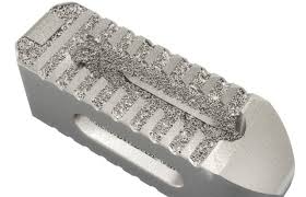
The Tritanium PL Cage is manufactured with Stryker’s innovative additive manufacturing process, also known as 3D printing. The cage is constructed using Stryker’s Spine division proprietary Tritanium technology, a novel, highly porous titanium alloy material designed for bone in-growth and biological fixation.1 Stryker’s 3D-printing process allows for the creation of porous structures that are designed to mimic cancellous bone, a type of spongy bone tissue.
“We are pleased to bring this technology advancement to spine surgeons and their patients,” said Stryker’s Spine division President Brad Paddock. “Stryker is a pioneer in 3D additive manufacturing, investing nearly 15 years in research and development. Unlike traditional manufacturing techniques, the flexibility of our 3D additive manufacturing capabilities allows us to precisely engineer and produce porous Tritanium devices. The Tritanium PL Cage is an exciting addition to our growing suite of unique spinal products.”
“Spine surgeons need a cage that has the capability of bony integration or bony in-growth, as well as radiolucency so that we can evaluate the fusion long term,” said Dr. Wellington Hsu, M.D., Orthopaedic Surgeon at Northwestern Medical Group. “Because Tritanium has favorable radiographic capabilities, as well as the integrative surface technology, that really in my opinion is what I would ask for from an interbody cage.”
Implanted via a posterior approach, the Tritanium PL Cage is available in a variety of widths, lengths, heights, and lordotic angles that can adapt to a variety of patient anatomies. Its large lateral windows and open architecture allow visualization of fusion on CT and X-ray, and its solid-tipped, precisely angled serrations are designed to allow for bidirectional fixation and to maximize surface area for endplate contact with the cage. The Tritanium PL Cage also is designed to minimize subsidence into the endplates.
The Tritanium PL Cage, which is produced at Stryker’s state-of-the-art 3D additive manufacturing facility, will be widely available to orthopaedic and neurosurgeons in mid-2016.
Tritanium Technology
In lumbar spinal fusion procedures, advancement of bony fusion at the target levels is at the cornerstone of a successful clinical outcome. In an effort to enhance the bony in-growth potential of implants, the scientific community has focused on porous metal implants in the hope of establishing a material similar in structure and mechanical properties to bone. Studies also have sought to understand which geometry and pore size would provide an optimal environment for cells to attach and multiply within this structure.2–4
Stryker’s Spine division conducted a pre-clinical animal study to investigate the biomechanical performance and bone in-growth potential of various lumbar interbody fusion implants utilizing different surface technologies (including the Tritanium PL Cage). The study has been accepted as a podium presentation at the NASS Annual Meeting being held Oct. 26-29, 2016, in Boston.
Indications for Use
The Tritanium PL Cage is indicated for use with autograft and/or allogenic bone graft comprised of cancellous and/or corticocancellous bone graft when used as an adjunct to fusion in patients with degenerative disc disease at one level or two contiguous levels from L2 to S1. Degenerative disc disease is back pain of discogenic origin with degeneration of the disc confirmed by history and radiographic studies. The degenerative disc disease patients may also have up to Grade I spondylolisthesis at the involved level(s). These patients should be skeletally mature and have six months of nonoperative therapy. Additionally, the Tritanium PL Cage may be used as an adjunct to fusion in patients diagnosed with degenerative scoliosis. It is to be implanted via a posterior approach and is intended to be used with supplemental spinal fixation systems that have been cleared for use in the lumbosacral spine.
About Stryker
Stryker is one of the world’s leading medical technology companies and, together with our customers, we are driven to make healthcare better. The Company offers a diverse array of innovative products and services in Orthopaedics, Medical and Surgical, and Neurotechnology and Spine that help improve patient and hospital outcomes. Stryker is active in over 100 countries around the world. Please contact us for more information at www.stryker.com.
Editor’s note: For images, video footage, or animation of Tritanium products and Stryker’s 3D additive manufacturing process, contact Barbara Sullivan at 714/374-6174 or [email protected]. A backgrounder about Tritanium and the Tritanium PL Cage also is available.
References
- Project #43909: Tritanium Technology Claim Support.
- Bobyn JD, Pilliar RM, Cameron HU, Weatherly GC. (1980) The optimum pore size for the fixation of porous-surfaced metal implants by the ingrowth of bone. Clinical Orthopaedics and Related Research, 150, 263-270.
- Webster TJ, Ejiofor JU. (2004) Increased osteoblast adhesion on nanophase metals; Ti, Ti6AI4V, and CoCrMo. Biomaterials, 25, 4731-4739.
- Karageorgiou V, Kaplan D. (2005) Porosity of 3D biomaterial scaffolds and osteogenesis. Biomaterials, 26, 5474-5491.
Content ID TRITA-PR-3
Contacts
Sullivan & Associates
Barbara Sullivan, 714/374-6174
[email protected]